65 Labeled Animal Cell Under Electron Microscope
To use a light microscope to examine animal or plant. It is an electron micrograph of cells largest and most important organelle the mitochondria and is characterized by the following features Fig.

Electron Microscopes Cell Structure Edexcel Gcse Combined Science Revision Edexcel Bbc Bitesize
Diagram Of Animal Cell Under Electron Microscope.
Labeled animal cell under electron microscope. Its a thin slice. E Material used in the course of Microscopic Anatomy at the University of Mainz. Labels are a means of identifying a product or container through a piece of fabric paper metal or plastic film onto which information about them is printed.
Investigating cells with a light microscope. Plant Cell Under Microscope Labeled. Draw and label an animal cell as seen under an electron microscope.
Wide collections of all kinds of labels pictures online. So lets begin by drawing a rough-oval shape. For example something that you draw as 3cm long may in fact be 10 000 times smaller in real life.
Labels are a means of identifying a product or container through a piece of fabric paper metal or plastic film onto which information about them is. We all do not forget that the human physique is quite problematic and a method I. Aims of the experiment.
Organelles In Plant And Animal Cells Venn Diagram Hamle Rsd7 Org. Now the first thing to point out when looking at images under an electron microscope is the scale. Make your work easier by using a label.
A Release of energy b Protein synthesis c Transmission of hereditary characters from parents to their off springs. So it is important to note that what we are drawing is definitely not life size. Posted 6 years ago.
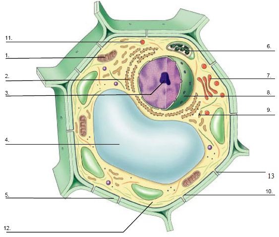
Both the global and high-resolution distribution of colloidal gold labels on cells can be readily determined. Draw a labelled diagram of the internal structure of a plant cell as seen with an electron microscope. Illustrate Only A Plant Cell As Seen Under Electron Microscope.
Animal Cells Under Microscope Labeled Written By MacPride Thursday January 11 2018 Add Comment Edit. Cell Wall Membrane Bacteriophage Black White Version Science Pics. Heres a diagram of a plant cell.
Cell is a tiny structure and functional unit of a living organism containing various parts known as organelles. Most cells both animal and plant range in size between 1 and 100 micrometers and are thus visible only with the aid of a microscope. 1 The name mitochondria was given by Benda 1898 and their ma n function was brought to light by Kingsbury 1912.
Diagram Of Animal Cell Under Electron Microscope Labeled. Learn the structure of animal cell and plant cell under light microscope. Some of these differences can be clearly understood when the cells are examined under an electron microscope.
Wide collections of all kinds of labels pictures online. In the given figure of an animal cell as observed under an electron microscope. Monday April 5th 2021.
Ii Which parts are concerned with the following functions. Animal Plant Cells Gcse Science Biology Get To Know Science Youtube Mitochondrion are visible with a light microscope but cant be seen in detail. Table D leads to images of electron microscopes or protocols for tissue preparation.
Blood opens an overview page on the different blood cells. Labeled Animal Cell Under Electron Microscope. Make your work easier by using a label.
Specialized cells that formed nerves and musclestissues impossible for plants to evolvegave these organisms mobility. Describe why it is important to prepare. These are both specific types of.
Iii Mention any two structures found only in plant cell not in animal cell. 2 Each mitochondria in section appears as sausage or cup or bowl shaped structure lined by double membranes. Thus light microscopes allow one to visualize cells and their larger components such as nuclei nucleoli secretory granules lysosomes and.
Label the cell wall cell-surface membrane capsule circular DNA flagella and plasmid. The limit of resolution of the light microscope is 02 µm while the practical limit of resolution of the electron microscope is about 1 nanometer nm. The diagram is very clear and labeled.
Ribosomes are only visible with an electron. A brief explanation of the. The animal cell is more fluid or elastic or malleable in structure.
The information can be in the form of hand-written or printed text or. I Name the parts labelled as 1 to 10. The lack of a rigid cell wall allowed animals to develop a greater diversity of cell types tissues and organs.
Here is an electron micrograph of an animal cell with the labels superimposed. Labelled animal cell diagram gcse. Photo Album By Darcy Plant And Animal Cells Under The Microscope.
Even though plant and animal cells are eukaryotic and share a few cell organelles plant cells are quite distinct when compared to animal cells as they perform different functions. But at the same time it is interpretive. Draw a labelled diagram of the internal structure of an animal cell as seen with an electron microscope.
Once slides have been prepared they can be examined under a microscope. Under the intense radiation of the electron microscope 011 electron per Å 2 the question of viability of cells naturally arises because the amount of radiation absorbed during highmagnification imaging is sufficient to cause cell death. Virtual Microscope Animal And Plant Cells Directions 1.
Ziehen die pins an die richtige stelle auf dem bild. You see that many features are in common. However no obvious structural damage.
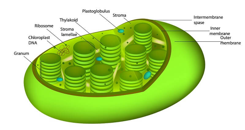
The plant cell as more rigid and stiff walls. Labeled Animal Cell Under Electron Microscope Chloroplast 145 Best Art Science Images On Pinterest Endoplasmic Reticulum Stock S Vectors Plant Stem Section Under the Microscope Detail. Labeled animal cell under electron microscope 8745961 orig.
So lets begin by drawing a rough-oval shape however this. Module 5 Page 2. Fillable Online 31 Name Date Diagram For Labelling Parts Of.
Monster Designs Animal Cell Under An Electron Microscope. See how a generalized structure of an animal cell and plant cell look with labeled diagrams.
32+ Plant And Animal Cell Structure Diagram
Animal cells Animal cell diagram. These are specialised for particular.
Plant Cell The Definitive Guide Biology Dictionary
Learn vocabulary terms and more with flashcards games and other study tools.
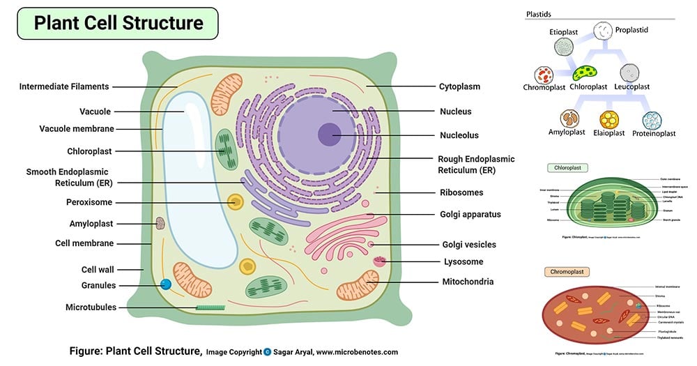
Plant and animal cell structure diagram. Plant Cell Diagram Educake. Plant cells have a cell wall and animal cells do not. Builds and transports substances through the cell.
Click on a video about plant cells and compare it to the video about animal cells. The animal cell diagram is widely asked in Class 10 and 12 examinations and is beneficial to understand the structure and functions of an animal. The most important structures of plant and animal cells are shown in the diagrams below which provide a clear illustration of how much these cells have in common.
Im in teaching profession since 2008. Breaks down food to produce energy in the form of ATP. Plant Cell And Animal Cell Diagram 8th Standard.
Learn vocabulary terms and more with flashcards games and other study tools. Nucleolus A round structure in the nucleus that makes ribosomes. Lets begin with the components of the animal cells-.
Teaching is my profession. Plant cells have chloroplasts which gives plants a green color. In both animals and plants cells generally become specialized to perform certain functions.
Generally plant cells are a lot bigger than the animal cells coming in more similar sizes and they are typically cubed or rectangular in shape. Bacteria fungi and some protists also have cell walls. Nucleus Cell membrane Cytoplasm Vacuole Cell wall Mitochondria Ribosomes Chloroplasts Animal cell Plant cell.
Nerve cells bone cells and liver cells for example all develop in ways that enable them to better perform their specific duties. My name is SANDEEP SINGH. Animal cells and plant cells are similar in that they are both eukaryotic cellsthese cells have a true nucleus which houses dna and is separated from other cellular structures by a nuclear membrane.
Education Chart Of Biology For Animal And Plant Cell Diagram. Now im using you t. Cell Wall Cell Membrane Nucleus Vacuole Chloroplast Plant and bacterial cell walls provide structure and protection.
The Plant Cell is the basic structural and functional unit found in the members of the kingdom Plantae. A bacteria diagram clearly helps us to profit extra approximately this unmarried cell organisms which have neither membrane-bounded nucleolus or organelles like mitochondria and chloroplasts. On this page we will learn about what is a plant cell definition structure model labeled plant cell diagram its cell organelles and the difference between plant cell and animal cell.
Plant and Animal Cell Structure - Labelled diagram. Plant cells also have structural organelles that are not found in the animals cells including the cell. 7 rows Animal cells usually have an irregular shape and plant cells usually have a regular shape.
29 rows Diagram showing the Difference between Plant cell and Animal cell As stated above both. In addition they have following organelles. Plant Animal Cell Diagrams and Graphic Organizer Unit 410 Handout 2 Extra WorkHomework Unit 410 Handout 3 6-way Paragraphs Introductory Level 35 pages 70 71.
Plant cells have all the structure mentioned in the animal cell except centrosome. Start studying Nielsen - Plant and animal cells. The cell wall is not a feature unique to plants.
They are different from plant cells in that they do contain cell walls and chloroplast. Animal cells do not have chloroplasts. Nucleus The control center of the cell.
The ultrastructure of a cell is its fine structure as revealed at high magnification. The major differences between plant and animal cells are. Golgi Body Processes and packages materials for the cell.
Contains the DNA Nuclear Membrane Surrounds the nucleus. Animal cells do not have cell walls. DEEPTHECHEMISTHello dear students and parents.
Vacuole Stores food and water. The structure labeled G give rise to spindle fibers and exclusively seen in animal cell. Structure of Plant cell.
Animal fungal and plant cells all contain structures called organelles. A plant cell is enclosed not only by the plasma membrane but also by a wall called the cell wall. Animal cells are eukaryotic cells that contain a membrane-bound nucleus.
